|
PhIFI Core
Facility (Phosphor-Imaging/Fluorescence Imaging) Brody 5S-21 |
|

|
PhIFI Core
Facility (Phosphor-Imaging/Fluorescence Imaging) Brody 5S-21 |
|
| |
Administrator: Dr. Brett D. Keiper contact: keiperb@ecu.edu |
|
| Brody School of Medicine/ECU
was awarded grants to set up a Core Facility for 2D imaging of chemiliumscent,
fluorescent and radioactive gel data, tissue sections and microarray data.
The Phosphor-Imaging/Fluorescence Imaging (PhIFI) core facility is available for use by all research laboratories on the East Campus and Brody Medical School of ECU. Labs must register annually with the Division of Research and Graduate Studies Office. PhIFI is located on the 5th floor in the Brody Building in the Department of Biochemistry and Molecular Biology in Room 5S-21. |

|
|
| Examples of Routine Typhoon Imager Uses: • Radioactivity detection from a blot or dried gel (32P,33P, 35S, 14C, etc.; requires Phosphorimage screen) • Chemiluminescent/Fluorescent detection on western blots using ECL+ • Multiple fluorophore labeled proteins, individual detection (e.g. Cy3 vs Cy5) • Fluorescent tag labeled DNA detection (e.g. ROX, TAMRA, etc.) on wet gels between plates • Imaging of slide-mounted DNA or Protein microarrays • Protein detection (in gel) by Sypro Ruby stain |

|
| Typhoon
Imager |
Registration |
ImageQuant
Software |
Technical Info |
Workstations |
Amersham/GE Healthcare |
|
Grant Funding from the North Carolina Biotechnology Center (NCBC) and the Division of Research and Graduate Studies was used to purchase a fully equipped Amersham/ GE Healthcare Typhoon 9410 Imager. |

|
East Carolina University
Brett D. Keiper, Ph.D. (Administrator) Dept of Biochemistry & Molecular Biology Brody School of Medicine Greenville, NC 27858 keiperb@ecu.edu |
Enhui Hao (Research Asst. III) Dept of Biochemistry & Molecular Biology Brody School of Medicine Greenville, NC 27858 Tel. 744-2693 haoe@ecu.edu |
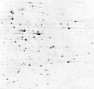
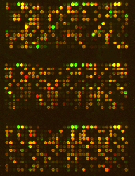
The above images are from the Amersham/GE Website:
(www5.amershambiosciences.com) |